BIBLIOGRAPHY
Forest, Herman Silva [Print] Handbook of Algae. The University of Tennessee Press. 1954
Thorp, and Covich [Print] Ecology and Classification of North American Freshwater Invertebrates. ed.3 Elsevier. Academic Press 2010.
McFarland, Kenneth [Internet] Botany 111 Fall 2014. [cited 11/17/2014].
Ost, and Rech. "Springs of Florida." Figure 1. Location of Selected Springs in Florida. Characteristics of Springs pag. USGS. USGS, May 1995. Web. 20 Nov. 2014.
Patterson. D.J. [Print] Free-Living Freshwater Protozoa. ed.1 Manson Publishing, ASM Press. 2009
Monday, December 1, 2014
Final Observation
For my final observation of the aquarium I was expecting a few things: 1. that it would be difficult to find new organisms; and 2. that my aquarium could be taken over by algae at this point. Neither of these suspicions were correct.
Because of the large amount of thine that passed between the observation and the set up of the aquarium, there was an increase in the algae that I found but was not so much that I could still see plenty of other organisms.
1. Calothrix sp.
 this pom-pom like structure makes the algae easier to identify when the entire structure is visible, but because it is more difficult to see if the branches are tapered or how they are connected, it took a bit more work to identify this organism.
For my final observation of the aquarium I was expecting a few things: 1. that it would be difficult to find new organisms; and 2. that my aquarium could be taken over by algae at this point. Neither of these suspicions were correct.
Because of the large amount of thine that passed between the observation and the set up of the aquarium, there was an increase in the algae that I found but was not so much that I could still see plenty of other organisms.
1. Calothrix sp.
2. Cladaphora sp.
 this is a nice picture because you are able to see the branchings which helps to identify the organism.
3. Coleochaete sp.

4. Difflugia sp.


This is the last of the algae that I recorded. Surprisingly I did not have a hard time finding these other organisms:
This is my favorite picture because this isn't just one difflugia, but it is two going through sexual reproduction. The difflugia on the right has pushed its vacuole into the difflugia on the left. This creates a gamete in the difflugia on the left which will soon bring rise to several more difflugia. The vacuole to the difflugia on the right will soon return to its place and they will disconnect and search for more reproduction partners.
5. Nematoda
This Nematoda was not very mobile, but it was interesting how it slithered through the water.
6. Litocolla sp.
This organism was the last one that I recorded. It is quite interesting as to how it gets its energy. All of the spike like objects surrounding the membrane act almost like a spider web. Fast moving organisms collide with the spikes and get wedged between them. When this happens the membrane opens and secretes a vacuole that encumbers the prey and then draws it back into the membrane where the prey is turned into energy.
That is all that I have for my observations. I wish that I would have been able to observe more but sadly time is an issue. I am interested to see how long it takes for the aquarium to be taken over by algae since I can the the sudden increase of algae in the aquarium.
Observation 2
It has been one week since my last observation but it seems that I might have missed quite a few organisms in my first observation because this week I found a good number of new microorganisms. I have no new videos, but some nice pictures.
1. Litonotus sp.
 I was quite impressed with this picture. The ability to see the organelles and all of its makeup is quite fascinating to me. The most interesting part was being able to see it move. It expanded and collapsed to stretch both ends of its membrane for movement.
It has been one week since my last observation but it seems that I might have missed quite a few organisms in my first observation because this week I found a good number of new microorganisms. I have no new videos, but some nice pictures.
1. Litonotus sp.
2. Craspedacusta sp.
 This guy is also referred to, more commonly, as a fresh water jelly fish. I saw no movement with this organism.
3. Elcane Rotifer sp.
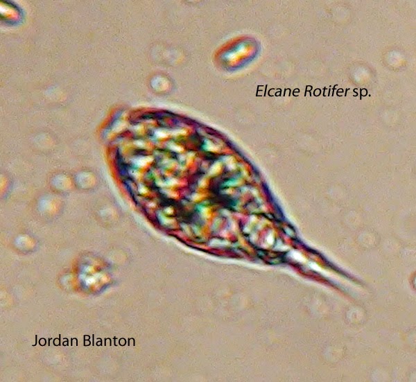 This organism seemed to be pretty abundant in my sample, it and other types of rotifers. You can see the bottom right hand corner of the picture is the flagella-like appendages that makes it mobile.
4. Paramicium sp.

All in all, this observation was a success. Like last time, there were a few organisms that I was not able to capture on camera but none of which were very abundant.
Observation 1
Sadly I was not able to get to my observations until now. My first one was 4 weeks after the set up. Because of the prolonged lack of observations and recordings I was not able to find the same microorganisms as I did when I first set up the micro aquarium, but that is not to say that this observation was unsuccessful. I was able to find many microorganisms that were bot spotted before. Also in my last post I left out my water source so for some clarification the source is from a spring that receives partial sun exposure in Carters Mill Park in Knoxville, Tennessee. So what did I find in the observation?
1. Amoeba sp. (seen above)
2. Limnius sp. (seen above)
3. Epalxis sp.
4. Vorticella sp.

5. Nodularia sp. (Forest, 1954).

Sadly I was not able to get to my observations until now. My first one was 4 weeks after the set up. Because of the prolonged lack of observations and recordings I was not able to find the same microorganisms as I did when I first set up the micro aquarium, but that is not to say that this observation was unsuccessful. I was able to find many microorganisms that were bot spotted before. Also in my last post I left out my water source so for some clarification the source is from a spring that receives partial sun exposure in Carters Mill Park in Knoxville, Tennessee. So what did I find in the observation?
3. Epalxis sp.
Along with these there were many fast moving microorganisms that were a bit too fast for me to catch on camera. I was also able to see Rotifer eggs and other signs of reproduction which tells me that there were suitable conditions for the thriving of the organisms within the water sample.
Monday, October 20, 2014
On October 14th I set up a micro aquarium form a water source in the Knoxville, Tennessee area. The specifics of the location are soon to come. Basically the procedure was taking a small, micro tank and and adding a bit of the sediment and water from different depths of the sample to ensure that the maximum amounts of organisms would be present in the observations. I then added two different plants to ensure that the organisms in the water were able to have the conditions needed the thrive for the extent of the observation process. More specifically the plants provided oxygen fixation through photosynthesis to sustain the microorganisms. Initially I observed three different types of micro organisms. One looked like a little bubble that moved by a spiral motion. Another looked like a worm wiggling for it's form of motion. And finally the one that seems most interesting to me was a large amount of single-celled organisms that attached themselves to one of the plants in the micro aquarium. The organism was triangular shaped and at the most acute point there was what resembled a string that came from the organism just like a spider dispels it's web. Once the organism got so far away from the plant while still attached by it's "web", it pulled itself in very quickly to bump into the other cells and near the plant it was attached to. I hypothesis that this is the way the organism gets energy but am not certain.
More information is soon to come and I will fill in the gaps that are currently in the post soon. I am excited to continue sharing the natural progress of this microsystem.
More information is soon to come and I will fill in the gaps that are currently in the post soon. I am excited to continue sharing the natural progress of this microsystem.
Subscribe to:
Posts (Atom)